|
 |
Trigeminal neuralgia
(Please wait while the video is downloading) |
Anatomy causes
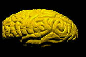
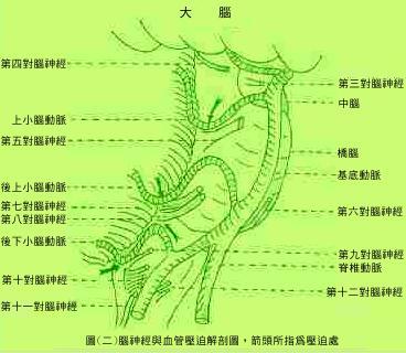
The trigeminal nerve is the thickest cranial nerve. After it is issued from the brain stem, it is divided into large sensory nerve roots and small motor nerve roots. Sensory nerveroots merge into a large ganglion after passing through the meninges, which is where the nerve cells are located. Then it was divided into three peripheral nerves, which were pierced through three small holes in the skull base and distributed to the face. The main function of trigeminal nerve is facial superficial sensation and its jurisdiction is shown in Figure 1. The etiology of typical trigeminal neuralgia is very clear, that is, the trigeminal nerve is compressed by artery or veins at the roots entry zone of brain stem ,then created a short circuit ,as shown in figure (b). This finding was discovered in the 1950s and 1970s by Prof. Dandy, Prof. Sunderlan,and finally proved by Prof. Petter Jannetta, a professor of Pisturburg University via a microvascular decompression procedure(MVD). Figure (c)
symptoms
Typical trigeminal neuralgia (TN)is unilateral and rarely seen on both sides. The second and third branches are more common. The pain attacked is sudden and severe,like a knife-cut,electric shock, which is lasting for few seconds,several hours to several days, then suddenly healed on its own and completely recovered without any sequale,but it will be attacked again soon. The longer the history of attack,the higher the recurrence rate. Some patients will experience "aura" before the attack, and always have " trigger point" (the starting point of severe pain). TN attacked usually When biting, eating cold food, brushing teeth or being nervous. Many patients suffer from pain from the gums. They are often mistaken for toothache and suffer from tooth extraction by dentists due to disdiagnosis.Even the entire row of teeth is uprooted but pain remains. There was a medical joke that 10,000 teeth in the United States have been removed due to misdiagnosed for trigeminal neuralgia each year. Patients and dentists should be pay attention to it and do not do it again.
diagnosis
Although symptoms of trigeminal neuralgia are obvious, they are still easily confused
Trigeminal neuralgia is characterized by:
1. It is a severe and sharp pain, not a soreness.
2. The trigger point (the starting point of the pain) is along the center of the face.
3. pain is entirely within the scope of the trigeminal nerve, never refered to neck and ear.
4. pain attacked and disappeared suddenly,if it occurs slowly or not supferficial,atypical trigeminal neuropathy is considered,which may be due to a tumor or inflammation.
5. Typical trigeminal neuralgia is not associated with other neurological disorders such as facial numbness, mscular weakness, difficulty swallowing or pain in the ear.
Differential diagnosis
1. Toothache: In terms of pain, both are severe tingling,but toothache is persistent, it is pain in the teeth and there will be no facial pain (touching the skin is not painful). Trigeminal neuralgia may be similar to toothache but will disappear suddenly without any sequalea.Trigeminal neuralgia can be diagnosed indirectally by drugs.
2. Tendonitis: It is caused by inflammation of the facial muscles due to improper use. It is a kind of soreness. It occurs in the tendon area, such as the TMJ syndrome. When the facial expression affects the tendon, it produces pain, such as laughing and eating hard food.
3. Geniculate neuralgia :
is a kind of neuralgia,patients have facial deep not superfical pain and had tenderness, and have ear pain.
4. atypical trigeminal neuralgia ( atypical TN):
although within the scope of the trigeminal nerve, but may be a numbness, tenderness, facial paresthesia,the firing point is not central, dramatic increase in the number of attacks, and so on.
5. facial pain of Unknown cause: pain is inconsistent and could be changing in locations or area.
treatment
With the right diagnosis, drug treatment or surgery is consdiered: Drug treatment is a palliative, and surgical treatment is a permanent cure.
1. Drug therapy: Generally, the patient receives medical treatment at the initial stage for one or two months.If it has good response then the surgery will be delayed, or the patients are too old or have meidcal probelms that is risky for anesthesia or surgery, durg therapy is continused.
2. Surgical treatment: since the TN caued bye vascular compression, MVD is very effecitve and sucessfull rate are more than 95%.
3. I am the only senior neurosurgeon in Taiwan who visited the University of Pittsburgh (1985) and followed Professor Jannetta in this field.I had at least 2000 cases received MVD surgical procedure.
Its complications with surgery
MVD is very delicate microsurgical procedure,the surgeon should be a good hand and a lot of expeerience.Otherwise it will cause quite a lot of complications, such as deafness, dizziness, tinnitus, if injury to the brain stem it leads to more
serious consequences. for acuumlating 30 years surgical experience my personal records are excellent not only the sequelaes are quite low, and only 3% of patients have temporary hearing loss.
Micrvascular Decompression
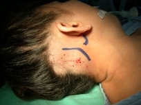
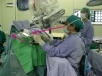
After the general anesthesia, a 3 cm skin incision was made behind the ear , and then a 1.5-cm skull defect was made by high speed drill, the dura opend and trigeminal nerve is identified ,then the vascular vessles are separated by insertion of teflon flet .The whole procedures usually finish within 60 minutes. (as shown below). After the operation, the patient need to staye in hospital for only 3-4 days. The pain usually completely disappeaed immidiately after operation, only a few neureous cases need take drugs agin for a couple of days.
Surgical wounds have been reduced to 3 cm in length,because of the accumulation of experience . It is located behind the ear and will not be seen after the operation due to hair cover.The head is fixed with three pin headrest.After general anesthesia.the whole surgical procedure is proceeding under the magnification of neurosurgical microscope.
By using a high-speed drill to remove a skull, a skull defect is made about 1.5 x 1 cm.the skull is backfilled after operation.
Surgery video :http://www.youtube.com/embed/ar5NY9xnQrE
|